
Translate this page into:
An unusual case of renal fungal mass masquerading as renal cell carcinoma
*For correspondence: vaibhavpedbhu@gmail.com
-
Received: ,
This is an open access journal, and articles are distributed under the terms of the Creative Commons Attribution-NonCommercial-ShareAlike 4.0 License, which allows others to remix, tweak, and build upon the work non-commercially, as long as appropriate credit is given and the new creations are licensed under the identical terms.
This article was originally published by Wolters Kluwer - Medknow and was migrated to Scientific Scholar after the change of Publisher.
A 12 yr old healthy male child† presented in the department of Pediatric Surgery, Institute of Medical Sciences, Banaras Hindu University, Varanasi, India, in January 2019, with dull-aching left flank pain for one month. Abdominal contrast enhanced computed tomography scan showed non-functioning left kidney with a mass and retroperitoneal lymphadenopathy, arising a suspicion of renal cell carcinoma (Fig. 1). A diagnosis of left renal fungal mass was made based on renal biopsy and urine wet mount (Figs 2 and 3). Following renal shut down on single intravenous liposomal amphotericin B, haemodialysis was done. The child showed improvement in pain and mass regression on itraconazole (10 mg/kg). On the seventh day after discharge, the child presented again with anuria. After haemodynamic stabilization, cystoscopy revealed urinary bladder filled with fungal colonies (Video 1). On laparotomy, fungal growth resembling a cake, encasing both the ureters and great vessels in the retroperitoneum was observed (Video 2). Despite these interventions, the child succumbed on the second postoperative day. This is rare case of progressive renal fungal infection in an immunocompetent child with extensive retroperitoneal spread.
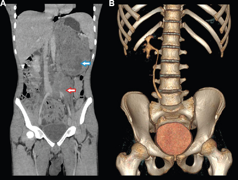
- (A) Contrast enhanced computed tomography of abdomen showing a left renal mass replacing the whole kidney (blue arrow) with retroperitoneal lymphadenopathy encasing left iliac vessels and abdominal aorta (red arrow) and (B) non-functioning left kidney on CT intravenous urography.
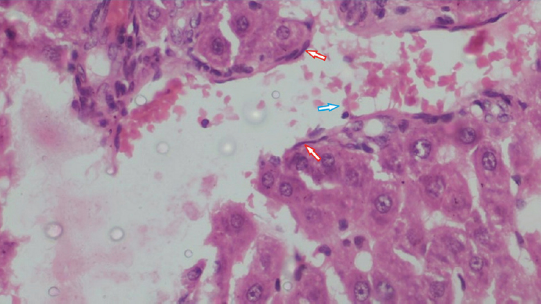
- Left renal biopsy showing the renal cortical cells with several aseptate fungal hyphae (red arrow) with dense inflammatory infiltrates comprising mainly neutrophils (blue arrow) (H&E stain; ×100).
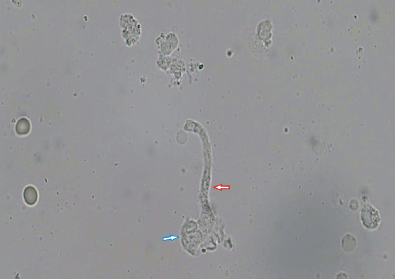
- Urine routine microscopy showing the aseptate fungal hyphae (red arrow) with macrophages (blue arrow) and red blood cells (×400).
 Videos available at ijmr.org.in.
Acknowledgment:
Authors acknowledge department of Radiology and Pathology, IMS, BHU for providing CT scan pictures and histology micro-photograph respectively.
Conflicts of Interest: None.


