
Translate this page into:
Coil mass extrusion from an embolized blownout carotid
*For correspondence: chiragkahuja@rediffmail.com
-
Received: ,
This is an open access journal, and articles are distributed under the terms of the Creative Commons Attribution-NonCommercial-ShareAlike 4.0 License, which allows others to remix, tweak, and build upon the work non-commercially, as long as appropriate credit is given and the new creations are licensed under the identical terms.
This article was originally published by Wolters Kluwer - Medknow and was migrated to Scientific Scholar after the change of Publisher.
A 35 yr old male†, with carcinoma of the tongue, previously treated with local excision and radiotherapy, presented to the Neurointervention Section of the department of Radiodiagnosis at the Postgraduate Institute of Medical Education & Research, Chandigarh, India, in September 2019, with a continuous bleed from a left-sided neck wound, resulting in haemodynamic shock. Computed tomographic (CT) angiogram revealed left carotid blowout (Figure A). Emergent digital subtraction angiography showed active contrast extravasation from the carotid (Figure B), which was immediately coil embolized (Figure C) after a crossflow evaluation. The patient was immediately revived and later discharged with no neurodeficits. After two weeks, he reported serous discharge from the neck wound and a hanging coil mass (Figure D). CT angiogram revealed partial coil mass exteriorization, however, with thrombosed left carotid artery (Figure E). The coil mass (Figure F) was carefully cut at the skin level. He survived for six weeks thereafter till he succumbed to tumour progression. This case highlights an unusual phenomenon of coil mass extrusion from a major superficially located embolized artery of the human body.
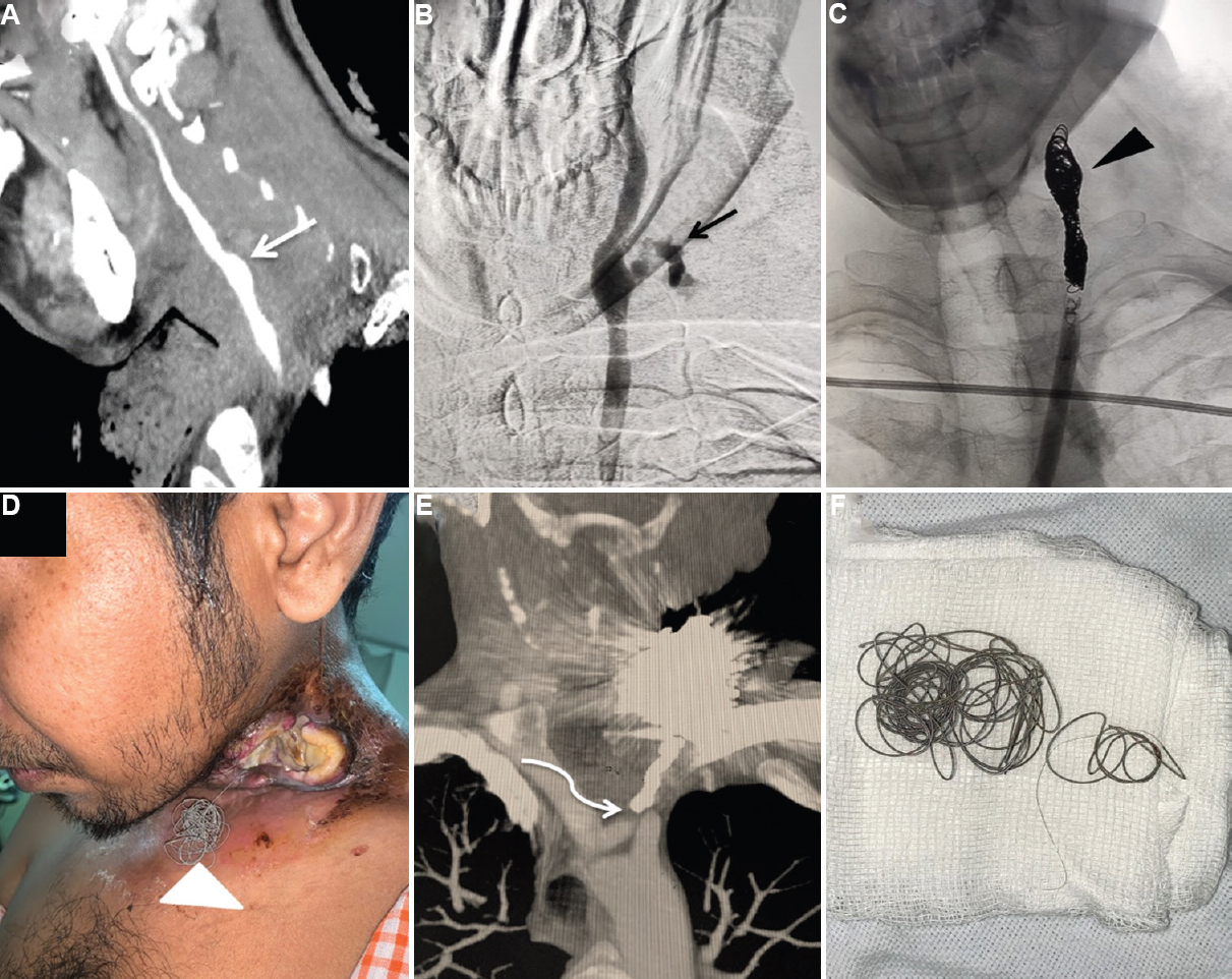
- Sagittal (A) maximum intensity projection computed tomography (CT) angiography image depicting focal dilatation of the carotid bulb (white arrow) confirmed on (B) catheter digital subtraction angiogram with active contrast extravasation suggestive of blowout (black arrow). Control digital subtraction angiogram image following coil embolization (C) showing coil mass (black arrowhead) in situ with cessation of the contrast efflux. Clinical photograph (D) showing hanging coil mass from the open neck wound (white arrowhead). (E) CT angiography depicting left common carotid thrombosis (curved arrow). (F) Actual extruded microcoil mass.
Conflicts of Interest: None.


