
Translate this page into:
Renal hydatid cyst involving inferior vena cava: A rare entity
*For correspondence: pankajzanwar.pz@gmail.com
-
Received: ,
This is an open access journal, and articles are distributed under the terms of the Creative Commons Attribution-NonCommercial-ShareAlike 4.0 License, which allows others to remix, tweak, and build upon the work non-commercially, as long as appropriate credit is given and the new creations are licensed under the identical terms.
This article was originally published by Wolters Kluwer - Medknow and was migrated to Scientific Scholar after the change of Publisher.
A 35 yr old female† presented to the Urology outpatient department at the King Edward Memorial Hospital, Mumbai, India, in June 2018, with a complaint of dull-aching right flank pain. On examination, the patient had palpable right renal lump of 8 × 6 cm in size. On computed tomography scan abdomen, the patient was diagnosed with right renal hydatid cyst with involvement of the inferior vena cava (IVC) (Figure A). The patient underwent right nephrectomy, with excision of around 5 cm length of IVC and reconstruction with a polytetrafluoroethylene graft with reimplantation of the left renal vein in the graft (Figure B-D). The patient was followed up till one year and was asymptomatic without recurrence.Venous invasion by renal hydatid cyst is a rare entity, and it may require complex reconstruction.
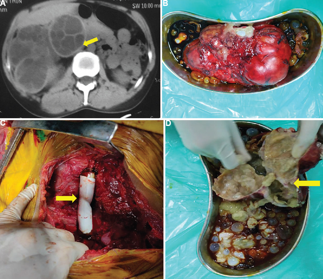
- (A) Computed tomography abdomen showing right renal hydatid cyst involving the inferior vena cava (arrow). (B) Excised specimen showing a large right renal hydatid cyst. (C) The inferior vena cava after reconstruction with polytetrafluoroethylene graft (arrow). (D) Specimen cut open to show the involved inferior vena cava wall (arrow) and daughter cysts (yellow arrow).
Acknowledgment:
Authors acknowledge the department of Cardiovascular and Thoracic Surgery, K.E.M. Hospital, Mumbai, Maharashtra, India, for providing vascular reconstruction images.
Conflicts of Interest: None.


