
Translate this page into:
Extensive palpebral molluscum contagiosum in an immunocompetent child
*For correspondence: prakash_2196@yahoo.co.in
-
Received: ,
This is an open access journal, and articles are distributed under the terms of the Creative Commons Attribution-NonCommercial-ShareAlike 4.0 License, which allows others to remix, tweak, and build upon the work non-commercially, as long as appropriate credit is given and the new creations are licensed under the identical terms.
This article was originally published by Wolters Kluwer - Medknow and was migrated to Scientific Scholar after the change of Publisher.
A seven year old boy† was brought to the Ophthalmology department at Lady Hardinge Medical College, New Delhi, India, in August 2017 with a six-month history of progressively enlarging raised lesions on his right upper and lower eyelids. On examination, he had three pitted globular lesions with central umbilication on the right upper eyelid and a similar lesion on the lower lid (Fig. 1) with the largest one being 9 mm × 8 mm × 6 mm with overlying prominent dilated vessel. Similar lesions were noted in the right nostril, the angle of mouth and ala of nose of the same side (Fig. 2). Complete blood count and immunoglobulin levels were normal. Serological tests for HIV and hepatitis A, B and C viruses were negative. CD4 count was not done. There was no history of the use of systemic steroids. Meticulous extirpation of the lesions was done along with cauterization of the base with trichloroacetic acid, with a good cosmetic outcome (Fig. 3). Histopathology showed intracytoplasmic eosinophilic inclusions (Henderson-Paterson bodies) within the keratinocytes of the basal, spinous and granular layers of epidermis, compressing the nucleus to periphery (Fig. 4), confirming molluscum contagiosum. No recurrence was noted at follow up after two months.

- Pitted globular lesions with central umbilication on the right upper and lower eyelid with overlying prominent dilated blood vessel.
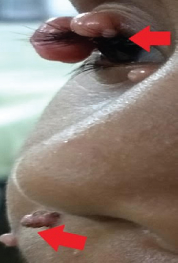
- Umbilicated lesions in the right nostril, angle of mouth and ala of nose.
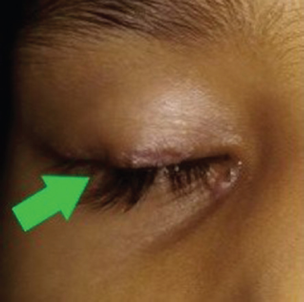
- Results after extirpation of the lesions along with cauterization of the base with trichloroacetic acid.
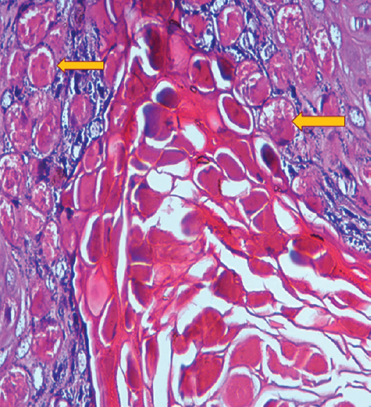
- Photomicrograph of the excised lesions showing intracytoplasmic eosinophilic inclusions (yellow arrows represent Henderson-Paterson bodies) within the keratinocytes, compressing the nucleus to periphery confirming molluscum contagiosum (H and E, ×40).
Molluscum contagiosum, a chronic infection caused by poxvirus in immunocompromised patients, is characterized by multiple centrally umbilicated papules. Treatment options include extirpation, cryotherapy, cauterization and topical cidofovir (5%) or imiquimod (5%) cream.
Acknowledgment:
Authors thank Dr Kiran Agarwal, Department of Pathology, Lady Hardinge Medical College, New Delhi, for kindly providing histopathology images and their interpretation that greatly improved the manuscript.
Conflicts of Interest: None.


