
Translate this page into:
Oculodermal melanocytosis
*For correspondence: shoryaazad@hotmail.com
-
Received: ,
This is an open access journal, and articles are distributed under the terms of the Creative Commons Attribution-NonCommercial-ShareAlike 4.0 License, which allows others to remix, tweak, and build upon the work non-commercially, as long as appropriate credit is given and the new creations are licensed under the identical terms.
This article was originally published by Wolters Kluwer - Medknow and was migrated to Scientific Scholar after the change of Publisher.
† Patient's consent obtained to publish clinical information and images
A 16 yr old male† presented to the Dr. Rajendra Prasad Centre for Ophthalmic Sciences, All India Institute of Medical Sciences (AIIMS), New Delhi, India, on August 21, 2017 with a complaint of discolouration around his eye. No history of systemic illness, trauma or drug intake could be elicited. His vision was 20/20 OU. His right-sided face and ocular surface had slate-grey to purple discolouration. Conjunctiva was freely mobile over immobile discolouration, suggestive of scleral pigmentation. His intraocular pressure was normal. Iris heterochromia was apparent OD (right eye) being darker (Fig. 1). Bruckner reflex and fundus photography showed dark-coloured fundus in OD (Figs 2 and 3). Diagnosis of oculodermal melanocytosis OD was made. The patient was put on observation, owing to propensity to develop glaucoma and risk of uveal melanoma. Oculodermal melanocytosis is a congenital disorder characterized by discolouration (slate-grey to purple) of skin, episclera, sclera, trabecular meshwork, uvea and orbital tissue due to increase in a number of melanocytes.
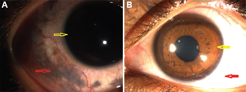
- (A) Right eye (OD) iris is darker (yellow arrow). Slate-grey discolouration of skin and sclera (red arrow). Conjunctival vessels show over pigmentation. (B) Normal pigmentation of other eye (OS) (yellow arrow over iris and red arrow over sclera).
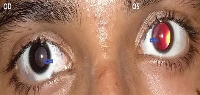
- Difference in Bruckner reflex of two eyes (blue arrow). OD glow is darker while OS glow is reddish-orange.
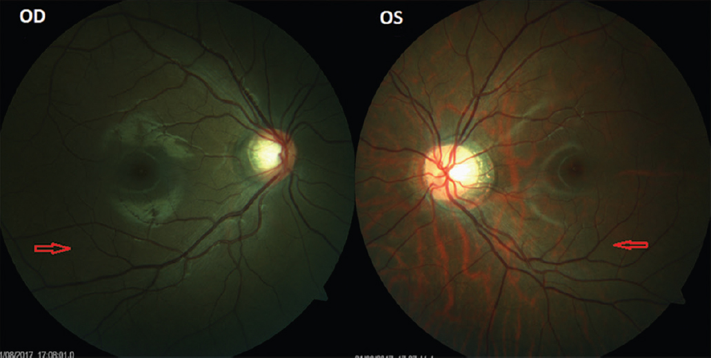
- OD fundus pigmentation is darker than OS (red arrow).
† Patient's consent obtained to publish clinical information and images
Conflicts of Interest: None.


