
Translate this page into:
Multisystem involvement of Langerhans cell histiocytosis in an adult
*For correspondence: nilaysengulsamanci@gmail.com
-
Received: ,
This is an open access journal, and articles are distributed under the terms of the Creative Commons Attribution-NonCommercial-ShareAlike 4.0 License, which allows others to remix, tweak, and build upon the work non-commercially, as long as appropriate credit is given and the new creations are licensed under the identical terms.
This article was originally published by Medknow Publications & Media Pvt Ltd and was migrated to Scientific Scholar after the change of Publisher.
A 27 yr old man† admitted to the Internal Medicine department of Haseki Training and Research Hospital, Turkey, in January 2015 with complaints of painful, itchy, red papules on his palms and soles for the past four months. He had diabetes insipidus in his medical history. Physical examination revealed diminished breath sounds and hepatomegaly. On his palms and soles erythematous-vesicle-squamous-crusty, lesions were seen (Fig. 1A). Aspartate aminotransferase: 587 U/l, alanine transaminase: 291 U/l, alkaline phosphatase (ALP): 696 U/l, gamma-glutamyl transferase (GGT): 1004 U/l, total bilirubin: 26 mg/dl, direct bilirubin:13 mg/dl were measured in the laboratory. Computed tomography findings reveal pleural thickening in the right hemithorax and bullous emphysematous lesions in both lungs, the largest measured 8 cm (Fig. 1B). Magnetic resonance cholangiopancreatography findings were compatible with sclerosing cholangitis. A tube was inserted endoscopically to the bile duct. The punch biopsies were taken from petechial lesions on the hands. Biopsy sections revealed epidermal hyperkeratosis with intra- and subepidermal pustules and at the base of pustules, histiocytes, eosinophilic infiltration of inflammatory cells containing polymorphs were seen. Histiocytic cells had a positive reaction with CD1a and it was consistent with the Langerhans cell histiocytosis group disease (Fig. 2A and B). After ursodeoxycholic acid and bile duct tube treatment, the value of the ALP, GGT and bilirubin decreased. The treatment consisted of systemic chemotherapy with vinblastine and prednisone. The patient died because of the respiratory failure.
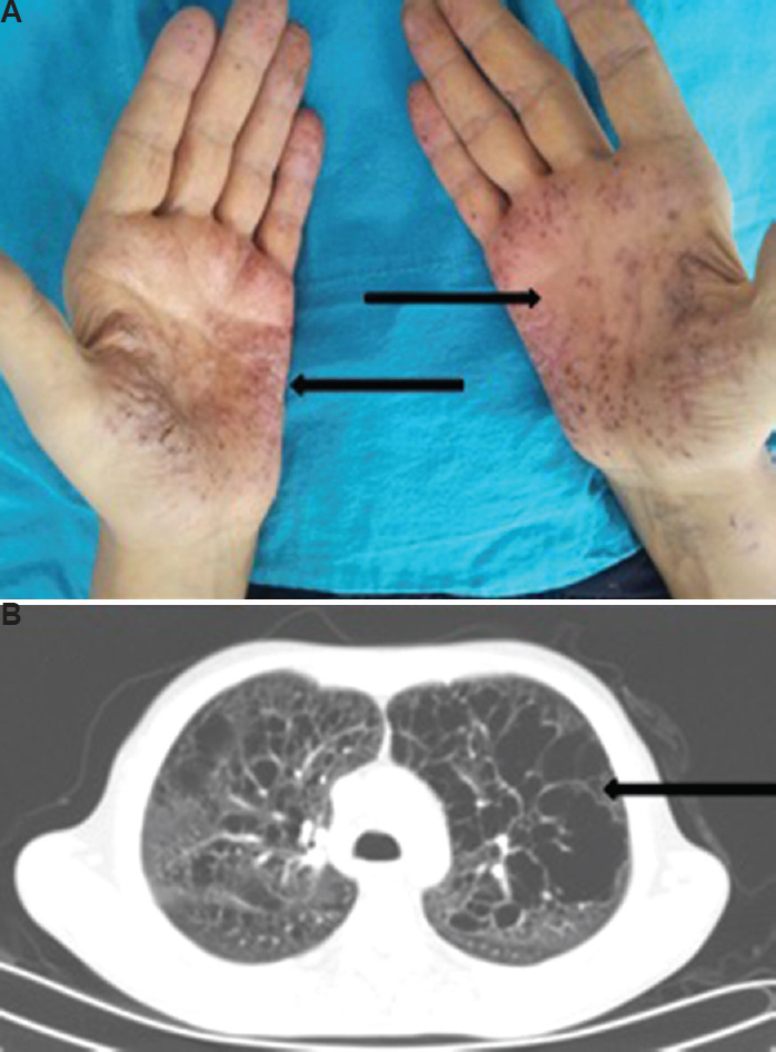
- (A) Erythematous-vesicle-squamous-crusty lesions on palms (arrows). (B) Computed tomography chest showing bullous emphysematous lesions in both lungs (arrows).
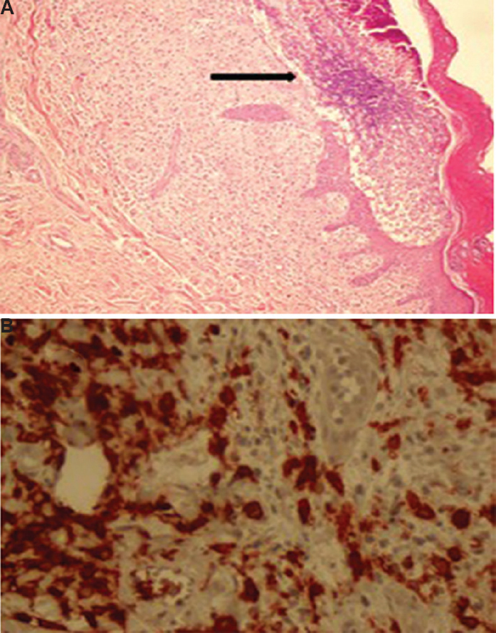
- (A) Epidermal hyperkeratosis with intra- and subepidermal pustules (arrow); and histiocytes, eosinophilic infiltration of inflammatory cells containing polymorphs and congested capillaries with extravasated red blood cell (H and E, ×100). (B) Positive immunohistochemistry for antigen CD1a (H and E, ×CD1a, ×400).
Acknowledgment
Authors thank Saime Gül Barut for histological assessment.
Conflicts of Interest: None.


