
Translate this page into:
Giant pleomorphic adenoma of submandibular gland
*For correspondence: sladjana-petrovic@hotmail.com
-
Received: ,
This is an open access article distributed under the terms of the Creative Commons Attribution NonCommercial ShareAlike 3.0 License, which allows others to remix, tweak, and build upon the work non commercially, as long as the author is credited and the new creations are licensed under the identical terms.
This article was originally published by Medknow Publications & Media Pvt Ltd and was migrated to Scientific Scholar after the change of Publisher.
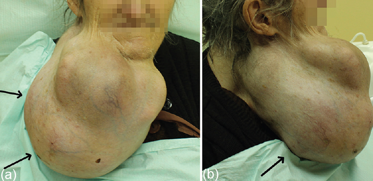
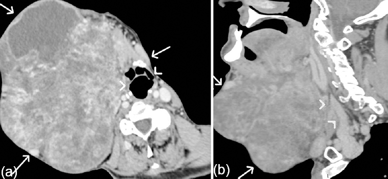
A 76 year old female patient presented to the department of Maxillofacial Surgery Dental Clinic in Nis, Serbia, in February 2014 with a large mass on the right side of the neck (Fig. 1a and b). The mass developed over the last 17 years. Physical examination showed pended, lobulated mass from the right submandibular region to the base of the neck. No cervical lymphadenopathy was found. Contrast-enhanced computed tomography revealed heterogeneous, lobulated contrast-enhancing lesion of 40 × 25 × 31 cm size (Fig. 2a and b). The lesion was characterized by the compression and displacement of the airway, carotid artery and internal jugular vein without infiltration (Fig. 2a and b).
-
(a) Patient with a pended, lobulated mass on the right side of the neck (arrows), frontal view. (b) Patient with a mass on the right side of the neck (arrows), lateral view.
-
(a) Axial post-contrast computed tomography scans showing heterogeneous, lobulated tumoural mass (arrows) with displacement of the airway (arrowheads). (b) Sagittal post-contrast computed tomography scans showing tumoural mass (arrows) with compression of internal jugular vein (arrowheads).
The patient underwent surgical therapy with complete surgical removal of the tumour mass weighting 3.5 kg. Histopathology showed submandibular gland pleomorphic adenoma composed of myxoid stroma and epithelial cells (Fig. 3). The follow up period was 18 months, with no evidence of recurrence (Fig. 4). This is a rare case of giant pleomorphic adenoma of submandibular gland with atypical location, long evolution without malignant alteration and complete surgical removal.
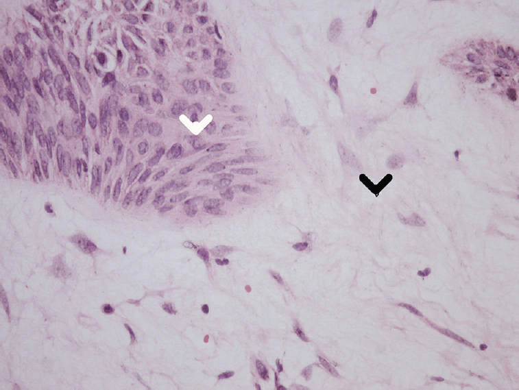
- Histopathology findings showed pleomorphic adenoma of submandibular gland composed of myxoid stroma (black arrowhead) and epithelial cells (white arrowhead) (Haematoxylin & Eosin, ×40).
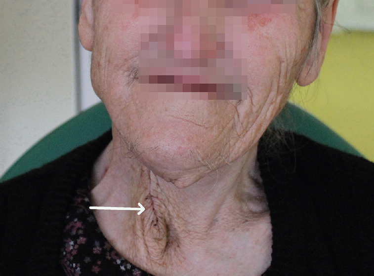
- Patient at six month follow up. Post-operative scar is present, with no evidence of recurrence (arrow).
Acknowledgment
Authors acknowledge the contribution of Dr Ivica Vucković, Department of Maxillofacial Surgery, Niš, Serbia.


