
Translate this page into:
Round pneumonia
* For correspondence: jclin@mail.ndmctsgh.edu.tw
This is an open access article distributed under the terms of the Creative Commons Attribution NonCommercial ShareAlike 3.0 License, which allows others to remix, tweak, and build upon the work non commercially, as long as the author is credited and the new creations are licensed under the identical terms.
This article was originally published by Medknow Publications & Media Pvt Ltd and was migrated to Scientific Scholar after the change of Publisher.
A 25 year old non-smoker male presented to the outpatient department of the Infectious Diseases Division, Kaohsiung Armed Forces General Hospital, Taiwan, in February 2014, owing to right anterior chest wall pain for three days. A posterior-anterior chest radiograph showed an opacification over right upper lobe (Fig. 1). Non-enhanced computed tomography demonstrated a 25mm nodule in the right upper lobe (Fig. 2). There was no lymphadenopathy. The patient was diagnosed to have round pneumonia and was treated with amoxicillin-clavulanate for seven days. Chest radiograph obtained five days later revealed a consolidation in the right upper lobe (Fig. 3). The sputum culture yielded Streptococcus pneumoniae. The blood culture was unremarkable. Repeat chest radiograph after 14 days showed regression of air-space opacification over right upper lobe (Fig. 4). Round pneumonia is usually seen in the children and is uncommon in adult.
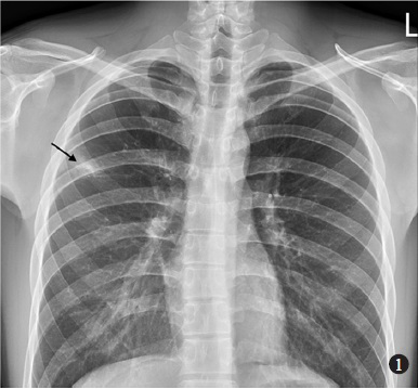
- Posterior-anterior chest radiograph showing opacification over right upper lobe (arrow).
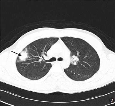
- Non-enhanced computed tomography demonstrated a 25mm nodule in the right upper lobe (arrow).
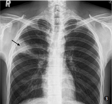
- Chest radiograph obtained 5 days later revealed a consolidation in the right upper lobe (arrow).
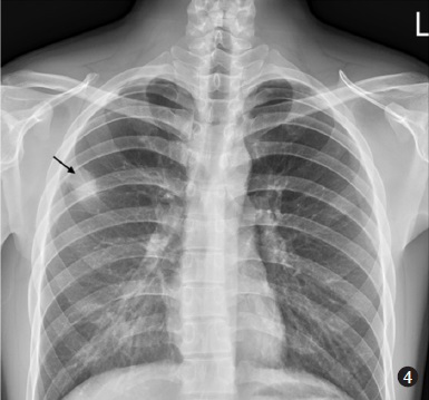
- Repeat chest radiograph after 14 days showed regression of air-space opacification over right upper lobe (arrow).
Acknowledgment
Authors thank Dr En-Han Huang, Department of Radiology, Kaohsiung Armed Forces General Hospital, Taiwan, for providing images.


