
Translate this page into:
Herpetic cheilitis
*For correspondence: kabirijdvl@gmail.com
This is an open access article distributed under the terms of the Creative Commons Attribution-NonCommercial-ShareAlike 3.0 License, which allows others to remix, tweak, and build upon the work non-commercially, as long as the author is credited and the new creations are licensed under the identical terms
This article was originally published by Medknow Publications & Media Pvt Ltd and was migrated to Scientific Scholar after the change of Publisher.
A 25 year old male presented to the out patient department of Dermatology, Lok Nayak Hospital, New Delhi, India, in April 2014 with fever and multiple pus filled lesions and raw oozy painful areas over the lower lip since last three days. There was gross swelling of lower lip. He had no history of similar complaints in the past. On examination diffuse swelling with eversion of lower lip was seen. A few pustules and multiple coalescing to discrete well defined tender ulcers of 0.5x 0.5 mm size with necrotic base and purulent discharge were present on the labial mucosa (Fig. 1A). Rest of the oral mucosa was normal. Regional lymphadenopathy was present. The patient had been treated with antibiotics and was advised to put Sumag (magnesium sulphate+urea) dressings from outside with no improvement. Though a possibility of contact dermatitis with secondary infection was considered, the erosions were suggestive of herpes and a Tzanck smear was made which revealed multinucleate giant cells admixed with neutrophils and lymphocytes (Fig. 2). HIV testing was negative. A diagnosis of herpetic cheilitis was made and the patient was started on tablet acyclovir 200mg five times a day with marked improvement in erosions and swelling in one week, (Fig. 1B) and with complete resolution of lesions in two weeks. This case represents a rare manifestation of a common disease.
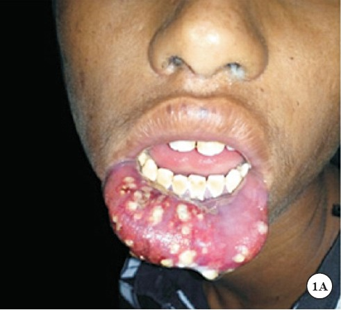
- (A)Photograph of the patient showing oedematous everted lower lip with a few pustules and multiple coalescing to discrete ulcers.
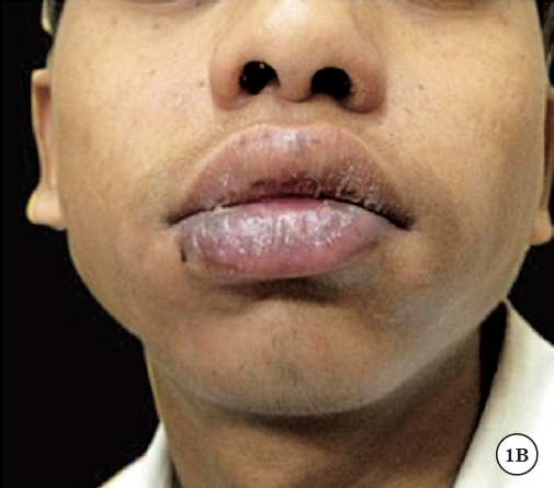
- (B)Marked improvement in erosions and swelling after one week of acyclovir therapy.
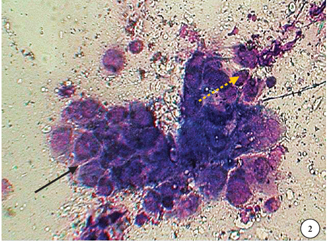
- Tzank smear made from the base of lesion showing multinucleate giant cell (black arrow) and a few secondary acantholytic cells (dotted arrow) at periphery (oil immersion field) × 100.


