Translate this page into:
Stewart-Treves syndrome
+For correspondence: stanczyk@poczta.onet.pl
This is an open-access article distributed under the terms of the Creative Commons Attribution-Noncommercial-Share Alike 3.0 Unported, which permits unrestricted use, distribution, and reproduction in any medium, provided the original work is properly cited.
This article was originally published by Medknow Publications & Media Pvt Ltd and was migrated to Scientific Scholar after the change of Publisher.
In November 2009, a 70-year old woman with chronic postmastectomy lymphoedema followed by chemotherapy and radiotherapy for invasive breast cancer, presented with 2 months history of enlarging plaques of purple nodules and massive bleeding from multiple ulcerating tumours of the left upper extremity (Figure). She was admitted in the department of General, Oncologic and Vascular Surgery of Military Health Service Institute in Warsaw, Poland. Biopsy confirmed clinical picture of angiosarcoma known as Stewart-Treves syndrome. Paliative amputation was performed of left arm. Though the wound healed well, she developed regional and distant metastases and died within a few months due to disseminated neoplasm.
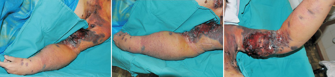
- (A, B, C). Clinical picture of Stevart-Treves syndrome: angiosarcoma arising within chronically oedematous skin of the left arm and forearm.
Stewart-Treves syndrome is a rare, deadly cutaneous angiosarcoma that develops in long-standing chronic lymphoedema. Though most commonly this angiosarcoma is a result of postmastectomy lymphoedema, it also develops in Milroy disease, idiopathic, congenital, traumatic and filarial lymphoedema.
The lesions typically appear as multiple reddish blue macules or nodules (Figure). Amputations or wide local excisions provide the best chance of long-term survival, chemotherapy and irradiation remain adjuvants to surgery. It is important to stress that the diagnosis of Stewart-Treves syndrome is usually established late because the inspection of chronically lymphoedematous skin with multiple hyperkeratotic nodules, fissures and papules and proper interpretation of clinical picture may be dificult. Biopsy is essential for the diagnosis of lymphangiosarcoma and should be performed to evaluate all suspicious skin lesions in patients with long-standing lymphoedema.


