Translate this page into:
Recurrent iris cyst following pencil tip injury
*For correspondence: ssnghy1@sify.com
This is an open-access article distributed under the terms of the Creative Commons Attribution-Noncommercial-Share Alike 3.0 Unported, which permits unrestricted use, distribution, and reproduction in any medium, provided the original work is properly cited.
This article was originally published by Medknow Publications & Media Pvt Ltd and was migrated to Scientific Scholar after the change of Publisher.
A 10 year old boy presented to Sri Sankaradeva Nethralaya, Guwahati, Assam, India, with history of injury with pencil tip in the right eye one month back and developed photophobia thereafter. Parents suspected retention of the broken pencil tip inside the eye. Slit lamp biomicroscopy revealed a transilluminant iris cyst on medial aspect of iris (Fig. 1, arrows). The cyst was excised twice but it recurred. On second recurrence, partial cyclectomy was done and in five years of post-operative follow up from March 2008 to November 2012 no recurrence was noted.
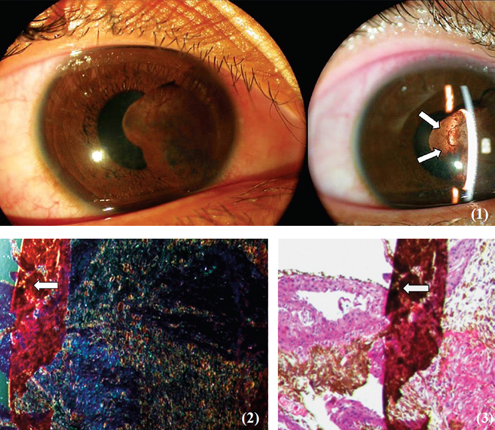
- Slit lamp biomicroscopy showing a transillu minant iris cyst on medial aspect of iris (arrows) (Fig. 1). Diffraction cytology (Fig. 2) and histopathological examination with Haematoxylin and Eosin stain of the excised tissue. (Fig. 3) showing presence of a foreign body (arrow in Figs 2 and 3×200).
Diffraction cytology (Fig. 2) and histopathological examination with Hematoxylin and Eosin stain of the excised tissue (Fig. 3) showed presence of a foreign body (arrow in Figs 2 and 3 × 200) suggesting that recurrence of iris cyst was a sequelae of retained pencil tip in the uveal tissue.
No further treatment was done as there was no reoccurrence and the pencil tip which was retained in the uveal tissue was removed by cyclectomy. The patients is under regular follow up.


