Translate this page into:
A study on inorganic elements in psammomas from ovarian & thyroid cancer
Reprint requests: Dr Eduardo Pérez-Campos, Biological & Medical Sciences Research Center, School of Medicine & Surgery, UABJO, Oaxaca, Mexico e-mail: perezcampos@prodigy.net.mx
-
Received: ,
This is an open-access article distributed under the terms of the Creative Commons Attribution-Noncommercial-Share Alike 3.0 Unported, which permits unrestricted use, distribution, and reproduction in any medium, provided the original work is properly cited.
This article was originally published by Medknow Publications & Media Pvt Ltd and was migrated to Scientific Scholar after the change of Publisher.
Abstract
Background & objectives:
Concentric lamellar calcifications known as psammoma bodies (PB) are found in benign and malignant tumours. Whether or not the inorganic element concentrations in psammomas are similar to serous adenocarcinoma of the ovary and thyroid papillary cancer tissues has not yet been ascertained. We undertook this retrospective study to establish if there is any difference in the concentrations of inorganic ions found in psammomas in serous adenocarcinoma of the ovary, and those found in thyroid papillary cancer tissue.
Methods:
PB samples from patients with adenocarcinoma of the ovary (n = 10) and with thyroid papillary cancer (n = 10) were analyzed through inductively-coupled plasma spectroscopy (ICP).
Results:
There were no significant differences in the concentrations of inorganic elements in PB from thyroid papillary cancer than in those PB from ovarian cancer.
Interpretation & conclusions:
Differences in the concentrations of inorganic elements may be due to the variation in environmental pollution. Our study had limitation of small sample size. Our results suggest that some inorganic elements can participate in the origin of psammoma bodies.
Keywords
Inductively coupled plasma
inorganic elements
psammoma
serous adenocarcinoma of the ovary
thyroid papillary cancer

Psammoma bodies (PBs) are concentrically laminated calcific spherules1–4. These are often observed surrounded by cells and frequently appear either acidophilic or basophilic on Papanicolaou stain with a concentric appearance. PBs usually are associated with papillary neoplasms of various organs as well as with a variety of benign conditions such as the use of intrauterine devices, oral contraceptives, endosalpingiosis, endometriosis, endometritis, thyroid and ovarian lesions and many others5–16. Earlier investigations have shown that patients age, sex, tumour size, histological grouping, extrathyroid invasion, and lymph node status were significant markers in predicting prognosis in patients suffering from papillary thyroid carcinoma17–19. Calcification (a frequent histological characteristic of papillary thyroid carcinoma) positively correlated with lymph node metastases of extra-thyroid invasion20.
Appearance of PBs is a well known histomorphological feature of ovarian adenocarcinomas. In some cases, to determine the origin of psammocarcinoma of the ovary requires the psammoma bodies to be present in at least 75 per cent of papillae which show a destructive invasion of the ovarian stroma21. The origin of psammoma bodies is not clear, but biomineralization has recently been found to be associated with a group of extremely small Gram-negative bacteria capable of precipitating calcium salts22, and in originating psammoma bodies. Moreover, the association of psammoma bodies with benign granulomatous reactions to a foreign material (aluminum silicate) has been reported23. Given that the presence of other inorganic elements in psammoma bodies is not known (and could be related to the activity concerning the metabolic regulation of cations) we decided to evaluate the participation of other inorganic elements in psammoma bodies, in ovarian and thyroid carcinomas. We performed a retrospective study of samples from patients with ovarian and thyroid cancer to compare the concentrations of inorganic elements among serous adenocarcinomas of ovarian and thyroid papillary cancers.
Material & Methods
The study was conducted between January 4, 2000 and March 1, 2010 in the Biological and Medical Sciences Research Center, School of Medicine and Surgery, UABJO, Oaxaca, Mexico. Inorganic elements from PB of serous adenocarcinomas of ovarian and thyroid papillary cancers were quantified by means of inductively-coupled plasma spectroscopy. Using a convenient sampling technique, paraffin-embedded tissues were selected from 20 male and female patients, (aged 45 to 77 yr) who had been diagnosed with either serous adenocarcinoma of the ovary, or thyroid papillary cancer. Cases were chosen from the archives of surgical pieces collected and kept by a pathologist since the year 2000.
All the surgical specimens were formaldehyde fixed, and paraffin embedded. These included 10 paraffin blocks each from serous adenocarcinoma of the ovary and thyroid papillary cancer. All paraffin-embedded tissue samples were confirmed by microscopy and contained PB. To prevent sample contamination by the analysts, paper face-masks and plastic gloves were worn during the preparation.
Paraffin blocks that met the criteria of calcified foci with concentric laminations located within the stromal stalks of tumour papillae of the thyroid were selected. Among the ovarian adenocarcinomas, round concentric laminations, (mostly intact structures with easily identifiable concentric laminations, and subjected to histological study with hematoxylin and eosin staining) were selected. All calcified masses that did not meet the criteria of psammoma bodies were discarded. Paraffin was removed from the blocks in a glass beaker at 60°C. The PB samples were selected, with a metallographic microscope with a 15X magnification (Olympus, USA). All psammoma bodies crystals were sifted through a 180-μm sieve. Crystals were dissolved in a CEM oven at 600°C (West Instrument, DGE-5825). For the analytical determinations of inorganic elements, a plasma emission spectrophotometer IRIS Intrepid II Inductively coupled plasma (ICP), model 460, Thermo Jarrell Ash from the Experimental Center of the Mexican Geological Service in Oaxaca, Mexico, was used, which was coupled to a plasma generation system of compressed argon gas at a pressure 80 psi, and a 99.99 per cent purity. Each sample was analyzed in duplicate. The aqueous standards calibration curve was prepared from a multi-standard solution. All solutions were prepared with deionized distilled and grade I water, with an electrical conductivity > 16.6 mW/cm2 at 25°C. Standards were prepared with certified reference high-purity materials at a micrograms per milliliter concentration in 5 per cent HNO324.
The ICP was calibrated to give a limit of detection for barium (Ba) 0.2, strontium (Sr) 0.1, phosphorus (P) 8, magnesium (Mg) 6, nickel (Ni) 0.3, sodium (Na) 7 and zinc (Zn) 0.1 in μg/g, and calcium (Ca) 1.4 per cent. Paraffin from blocks was used as negative controls in order to discard inorganic elements that could have affected the results. Blank corrections were not necessary.
The study and enrollment strategy were approved by the Institutional Review Board of the Master in Sciences Program of the Medical School of the UABJO, Oaxaca, Mexico. Before enrollment, written informed consent was obtained from all subjects.
The results were analyzed by Mann Whitney test, using GraphPad Prism version 5.00 for Windows, GraphPad Software, San Diego, CA, USA.
Results & Discussion
Because the inorganic elements in the PB showed asymmetrical frequency distributions, the geometric mean and minimum and maximum values were recorded for the inorganic elements. As a control, calcium was measured in paraffin blocks; using inductively-coupled plasma spectroscopy, calcium was less than 8.5 per cent in the paraffin utilized.
All PB from serous adenocarcinomas of ovarian and thyroid papillary cancers contained in their structure at least eight inorganic elements: barium, calcium, strontium, phosphorus, magnesium, nickel, sodium, and zinc (Table). Aluminum (Al), cadmium (Cd), cobalt (Co), copper (Cu), chromium (Cr), iron (Fe), and potassium (K) were also found in psammoma bodies from serous adenocarcinomas of the ovarian and thyroid papillary cancers. Statistical analysis showed no significant difference between the concentration of cations in the samples of psammoma bodies and the serous adenocarcinomas of ovarian and thyroid papillary cancers.
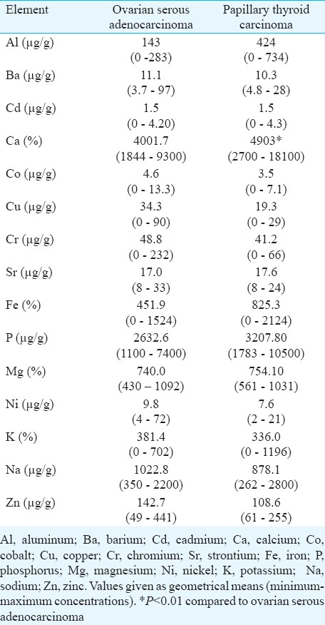
Inorganic element concentrations in PB from ovarian and thyroid cancer tissues are not reported. When inorganic element concentrations from PB of serous adenocarcinomas of the ovarian tissue were compared with those reported by Yaman et al25 from ovarian tissues with cancer it was observed that the PB had 12 times more copper (Cu); about 10 times more cadmium (Cd), calcium (Ca), iron (Fe), and about 5 times more magnesium (Mg) and zinc (Zn) than ovarian tissues. These increased concentrations in PB can be explained by modifications in the proteins that participate in the metabolic regulation of cations or environmental pollution.
The importance of looking for differences in the concentration of cations in PB from serous adenocarcinomas of ovarian and thyroid papillary cancers, especially calcium, is the presence of proteins like small calcium-binding proteins26; the proteins related to the PB, the bone morphogenetic proteins (BMPs) family, especially BMP-11, and the osteopontin produced by macrophages that is related to the development of PB in papillary carcinoma of the thyroid27, could explain the differences in cation concentration in PB.
Considering that the increased inorganic elements in PB may be related to the environment, we compared some of these elements reported in gallstones, and found that aluminum (Al), magnesium (Mg), and zinc (Zn) were found in higher concentration in PB28. This could suggest the presence of concentrating mechanisms, or environmental contamination. The identification of PB was made based on criteria of calcified foci with concentric laminations, discarding samples with stromal calcifications and bone formation. Moreover, the proportion of inorganic elements found in this study cast doubt on the hypothesis that PB are formed by successive layers where calcium salts were deposited29.
In conclusion, there were no differences in the concentrations of inorganic elements in psammomas from thyroid papillary and ovarian cancer, but our results suggest that other inorganic elements can participate in the origin of psammoma bodies. Further studies are required with a larger sample size to confirm or findings.
References
- Survival impact of psammoma body, stromal calcification, and bone formation in papillary thyroid carcinoma. Mod Pathol. 2009;22:887-94.
- [Google Scholar]
- Significance of psammoma bodies in serous cavity fluid: a cytopathologic analysis. Cancer. 2004;102:87-91.
- [Google Scholar]
- psammoma bodies- Friends or foes of the aging choroid plexus. Med Hypotheses. 2010;74:1017-20.
- [Google Scholar]
- Microcalcifications and psammoma bodies in thyroid tumors. Thyroid. 2008;18:1017-8.
- [Google Scholar]
- The significance of psammoma bodies in screening cervical cytologic smears. Am J Obstet Gynecol. 2003;188:1609-12.
- [Google Scholar]
- Psammoma bodies in papillary adenocarcinoma of the endocervix. Int J Gynecol Pathol. 1983;2:216-21.
- [Google Scholar]
- Cervicovaginal psammoma bodies in endosalpingiosis.A case report. J Reprod Med. 2000;45:526-8.
- [Google Scholar]
- Psammoma bodies of benign endometrial origin in cervicovaginal cytology. Acta Cytol. 1977;21:550-2.
- [Google Scholar]
- Psammoma bodies in the cervicovaginal smear in association with benign papillary structures of the ovary. Acta Cytol. 1970;14:45-7.
- [Google Scholar]
- Psammoma bodies in a cervicovaginal smear associated with an intrauterine device. A case report. J Reprod Med. 1987;32:147-8.
- [Google Scholar]
- Psammoma bodies and detached ciliary tufts in a cervicovaginal smear associated with benign ovarian cystadenofibroma. Acta Cytol. 1980;24:549-52.
- [Google Scholar]
- Psammoma bodies in a cervical smear in association with borderline ovarian epithelial malignancy. J Pak Med Assoc. 1998;48:52-3.
- [Google Scholar]
- Psammoma bodies in the cervicovaginal smear in association with a papillary tumor of the peritoneum. Obstet Gynecol. 1983;61:130-4.
- [Google Scholar]
- Non-tumor-associated psammoma bodies in the thyroid. Am J Clin Pathol. 2003;119:90-4.
- [Google Scholar]
- The origin and significance of thyroid psammoma bodies. Lab Invest. 1980;43:287-96.
- [Google Scholar]
- Papillary carcinoma. In: Delellis RA, Lloyd RV, Heitz PU, Eng C, eds. World Health Organization Classification of tumors, pathology and genetics of tumors of endocrine organs. Lyon, France: International Agency for Research on Cancer; 2004. p. :57-66.
- [Google Scholar]
- Papillary carcinoma of the thyroid in Japan: subclassification of common type and identification of low risk group. J Clin Pathol. 2004;57:1041-6.
- [Google Scholar]
- Subclassification of non-solid-type papillary thyroid carcinoma identification of high-risk group in common type. Cancer Sci. 2008;99:1908-15.
- [Google Scholar]
- The origin and significance of thyroid psammoma bodies. Lab Invest. 1980;43:287-96.
- [Google Scholar]
- Serous psammocarcinoma of the ovary: An unusual finding. Gynecol Oncol. 2005;99:510-1.
- [Google Scholar]
- Presence of nanobacteria in psammoma bodies of ovarian cancer: Evidence for pathogenetic role in intratumoral biomineralization. Histopathology. 2004;45:633-7.
- [Google Scholar]
- Aluminum silicate-containing psammoma bodies in a cervicovaginal smear (Pap): cytological, ultrastructural, and radiographic microprobe studies. Diagn Cytopathol. 1999;21:122-4.
- [Google Scholar]
- A new continuous calibration method for inductively coupled plasma spectrometry. Anal Bioanal Chem. 2006;384:531-41.
- [Google Scholar]
- Comparison of trace element concentrations in cancerous and noncancerous human endometrial and ovary tissues. Int J Gynecol Cancer. 2007;17:220-8.
- [Google Scholar]
- Expressión of S100A2 and S100A6 in thyroid carcinoma. Histopathology. 2005;46:569-75.
- [Google Scholar]
- Possible relation of osteopontin to development of psammoma bodies in human papillary thyroid cancer. Arch Pathol Lab Med. 1998;122:1087-90.
- [Google Scholar]
- Inductive coupled plasma spectroscopy study of trace elements in gallstones from different regions of Oaxaca State. Salud Publica Mex. 2005;47:191-2.
- [Google Scholar]
- Heterotopic ossification and psammomatous calcification in gastric carcinoma. Case report and review of the literature. Acta Pathol Jpn. 1979;29:975-84.
- [Google Scholar]


